Technology based on state-of-the-art academic research

Our technology is based on many years of research at the KTH Royal Institute of Technology in Stockholm, Sweden. We have condensed our experience of both accurate hardware control and careful image reconstruction into a high-performance product.

We use propagation-based phase-contrast tomography, since it has been proven to be the best way to obtain high-resolution images of low contrast samples. The principle of the setup is simple, using an x-ray source, the sample stage and a detector with free-space propagation distances in between. The price of the simple setup is paid by high demands on the equipment and more complex reconstruction. Our product is based on the Excillum MetalJet x-ray sources, since they provide stable operation and the highest possible brightness from a compact source.

Performance examples

An ancient mummified hand

Mummies and other ancient remains are commonly examined by x-ray imaging. Bone provides the strongest contrast and has been the main source of information. In paleopathology, the study of ancient diseases, significantly more information can be extracted if also studying the soft tissue at high resolution. Here, we show an ancient Egyptian mummy hand scanned with the Exciscope technology.

A. Axial view through tip of middle finger. B. Sagittal section. C. Adipose tissue with the remains of adipose cells. Scalebars represent 1 mm.


Carbon-fiber composites

The Exciscope technology is at its best when used for samples consisting of materials with low atomic numbers. These materials typically give low contrast in x-ray imaging, an obstacle that is overcome by using the Exciscope technology. This enables non-destructive testing with high detail of materials used for lightweight constructions. In this example, we have imaged the interior microstructures in the carbon-fiber composite shaft of a golf club. The images show clearly the material design, fiber orientations and internal voids.

Axial slice of the golf shaft depicting the construction from carbon fiber sheets and internal voids in the material.
Tangential slice of the golf shaft shows the different fiber orientations in the sheets.


Atherosclerosis in coronary arteries

The Exciscope technology is well suited for imaging biomedical tissue samples, in this example excised human coronary arteries. The purpose was to observe microscopic atherosclerotic plaques, both calcium-rich and lipid-rich. This work shows that it is possible to observe small lesions, down to single or a few foam cells large, in the complete three-dimensional sample. This example can be extended also to other tissue types, and proves the unique abilities of the Exciscope technology in three-dimensional imaging of small low-contrast features in centimeter-sized tissue samples.

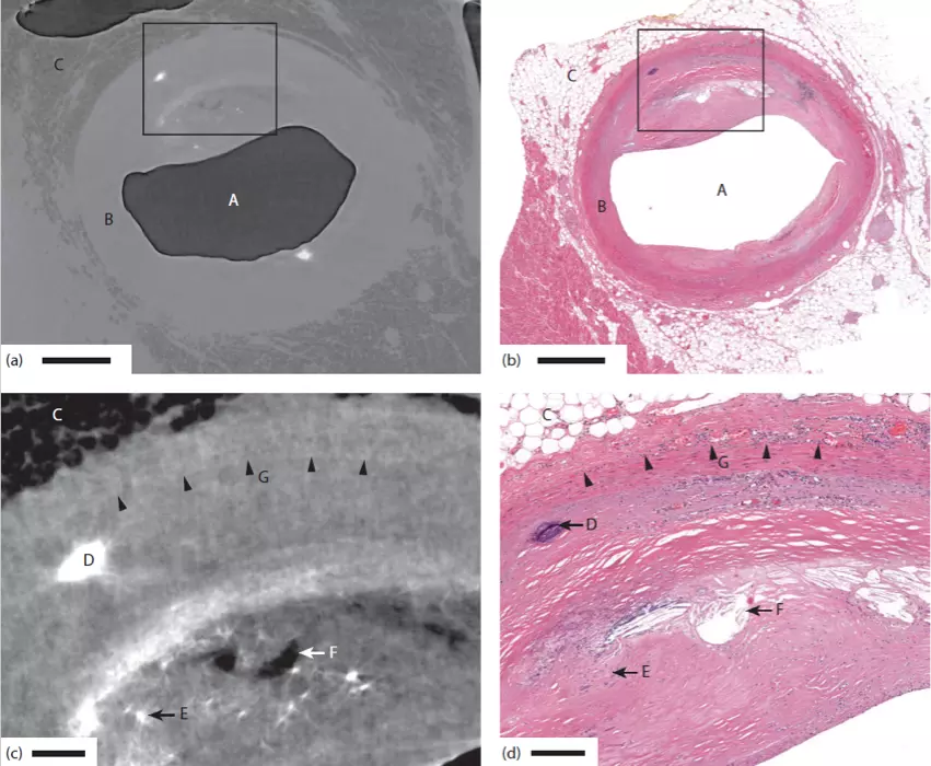
The figure depicts a comparison between the Exciscope 3D technology and conventional 2D histology. In the images are (A) air-filled artery lumen, (B) the artery wall, (C) adipose tissue, (D) calcification, (E) microcalcification, (F) cholesterol crystal depositions, and (G) demarcation between tunica media and tunica adventitia. Scale bar: (a,b) 1 mm, (c,d) 200 μm.

Reproduced (license) from W. Vågberg et al., “Cellular-resolution 3D virtual histology of human coronary arteries using x-ray phase tomography”, Scientific Reports (2018).


Zebrafish muscle imaging

As an example of the ability to use the Exciscope technology to image samples providing extremely low contrast, we show muscle myofibril imaging in whole-body zebrafish. In this application, healthy and dystrophin deficient zebrafish are compared, showing that this technology can be useful for physiological studies.

Phase-contrast tomography of juvenile zebrafish. (a) Axial slice, corresponding to the
dashed line through (b). Arrows indicate (from top) muscle tissue, notochord, and stomach. Scale bar is 100 μm. (b) Sagittal slice, corresponding to the dashed line through (a). The myofibril pattern is clearly visible. The arrows (from left) indicate bone, swim bladder and two myosepta. The scale bar is 100 μm. (c) Enlargement of the boxed area in (b). Scale bar is 50 μm.

Reproduced (license) from W. Vågberg et al., “X-ray phase-contrast tomography for high-spatial-resolution zebrafish muscle imaging”, Scientific Reports (2015).


Fast imaging of low-Z materials

Traditionally, fast high-resolution x-ray imaging has only been done at synchrotrons, since these sources provide very high brightness. However, synchrotron facilities are rare and expensive, so beamtime requires successful application, detailed planning and travelling. The Exciscope technology now enables some imaging applications to be done in the home lab, that were previously only possible at synchrotrons!

Fast phase-contrast image of a cheese doodle acquired in 75 milliseconds. The image shows structures down to 10 µm in the foamy structure based on carbohydrates and fat. Salt crystals appear dark in the x-ray image.

List of publications

  1. J. Romell et al., “Soft-Tissue Imaging in a Human Mummy: Propagation-based Phase-Contrast CT”, Radiology (2018).
  2. W. Vågberg et al., “Cellular-resolution 3D virtual histology of human coronary arteries using x-ray phase tomography”, Scientific Reports (2018).
  3. D. Larsson et al., “High-resolution short-exposure small-animal laboratory x-ray phase-contrast tomography”, Scientific Reports (2016).
  4. W. Vågberg et al., “X-ray phase-contrast tomography for high-spatial-resolution zebrafish muscle imaging”, Scientific Reports (2015).