As an example of the ability to use the Exciscope technology to image samples providing extremely low contrast, both whole-body imaging as well as 1 µm high-resolution imaging on the head of a zebrafish have been performed. In the high-resolution example, a comparison study of lab CT systems and a synchrotron source were completed, including the prototype version of the Exciscope instrument. In the whole-body application, healthy and dystrophin deficient zebrafish are compared, showing that this technology can be useful for physiological studies.
A
B
Föregående bild
Nästa bild
Phase contrast tomography of zebrafish head. (A) Reconstructed slice achieved by the TOMCAT beamline at SLS using a total scan time of 0.15 hrs. (B) Comparable reconstructed slice achieved by Exciscope Polaris using a total scan time of 6 hrs, demonstrating 1µm observable resolution of similar quality in a lab system.
Reproduced from A. Migga et al., “Comparative hard x-ray tomography for virtual histology of zebrafish larva, human tooth cementum, and porcine nerve”, Journal of Medical Imaging (2022).
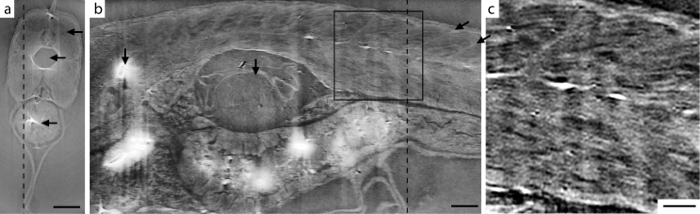
Phase-contrast tomography of juvenile zebrafish. (a) Axial slice, corresponding to the dashed line through (b). Arrows indicate (from top) muscle tissue, notochord, and stomach. Scale bar is 100 μm. (b) Sagittal slice, corresponding to the dashed line through (a). The myofibril pattern is clearly visible. The arrows (from left) indicate bone, swim bladder and two myosepta. The scale bar is 100 μm. (c) Enlargement of the boxed area in (b). Scale bar is 50 μm.
”Reproduced from W. Vågberg et al., “X-ray phase-contrast tomography for high-spatial-resolution zebrafish muscle imaging”, Scientific Reports (2015).
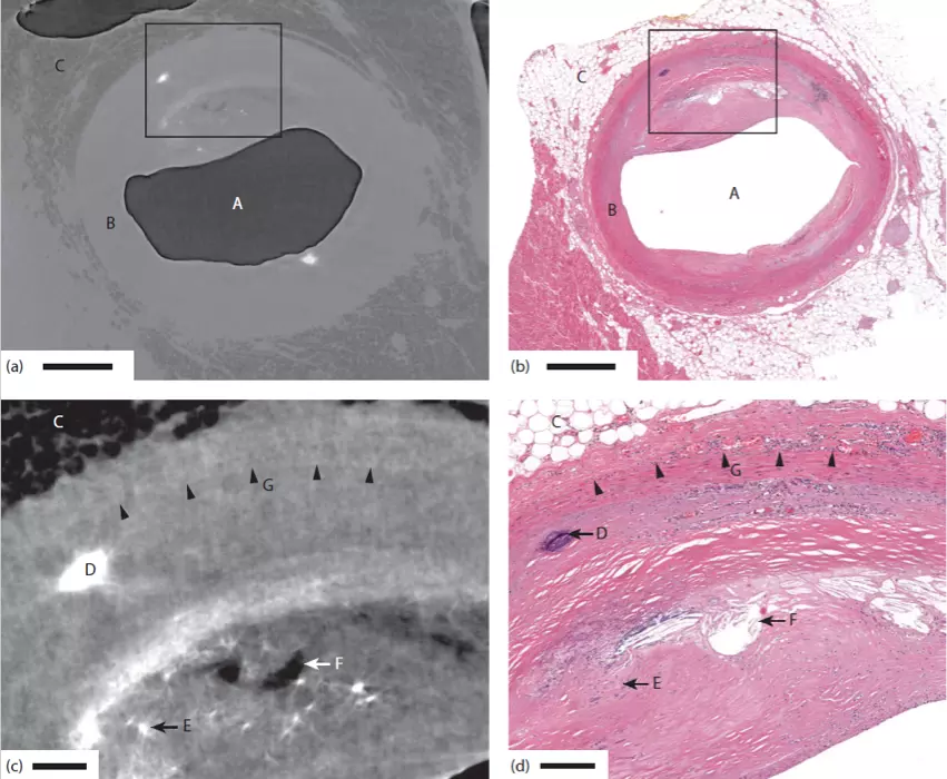
“Reproduced from W. Vågberg et al., “Cellular-resolution 3D virtual histology of human coronary arteries using x-ray phase tomography”, Scientific Reports (2018).”
The figure depicts a comparison between the Exciscope 3D technology and conventional 2D histology. In the images are (A) air-filled artery lumen, (B) the artery wall, (C) adipose tissue, (D) calcification, (E) microcalcification, (F) cholesterol crystal depositions, and (G) demarcation between tunica media and tunica adventitia. Scale bar: (a,b) 1 mm, (c,d) 200 μm.
Tracking veins and arteries in 3D
The Exciscope Polaris is well suited for imaging biomedical tissue samples, such as excised human coronary arteries. The purpose of the study was to observe microscopic atherosclerotic plaques, both calcium-rich and lipid-rich. This work shows that it is possible to observe small lesions, down to single or a few foam cells large, in the complete three-dimensional sample. This example can be extended also to other tissue types and proves the unique abilities of the Exciscope technology in three-dimensional imaging of small, low-contrast features in centimeter-sized tissue samples.