Tumors and tissues: a better way to study the human body
Surgery is an essential portion of the curative plan for patients with solid tumors. The outcome of these tumor surgeries (e.g. recurrence rates and patient survival) depends on several factors such as the resection margin. This is the amount of healthy tissue surrounding the tumor in the removed portion of tissue, demonstrating that the entire tumor has been removed. Therefore, the surgery is followed by a pathological assessment of the tumor itself, as well as the surrounding tissue. Standard histopathological analysis is a time-consuming process often ranging from weeks to a few days after the surgery at a minimum. The speed of the Exciscope Polaris, allows for faster assessment of the resection margins while simultaneously providing valuable 3D information.
Taking Histology from 2D into 3D
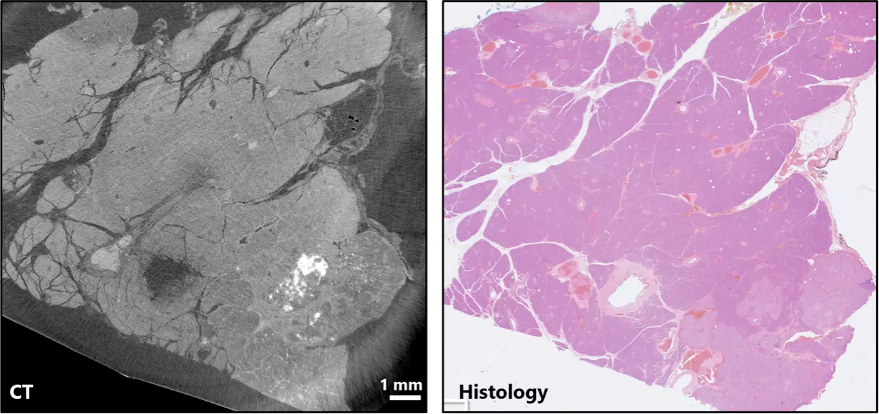
A comparison of unstained paraffin-embedded standard histological tissue samples from a biobank is shown below. Analysis of a pancreatic neuroendocrine tumor with the Exciscope Polaris and classical histology methods show similar quality, with the Exciscope Polaris highlighting the calcified scar tissue that is less visible in the histology analysis. This demonstrates that the Exciscope Polaris can be used as a nondestructive tool to complement a pathologists other histology techniques.
3D rendering of phase contrast CT data from the Exciscope Polaris of the neuroendocrine tumor pancreas sample highlighting the calcified scar tissue. Certain 2D slices could miss this feature entirely.
Reproduced (license) from W. Twengström et al., “Can laboratory x-ray virtual histology provide intraoperative 3D tumor resection margin assessment?”, Journal of Medical Imaging (2022).
Virtual slice (left) and histological slice (right) of approximately the same locations of a neuroendocrine tumor from a pancreas tissue sample.