Applications
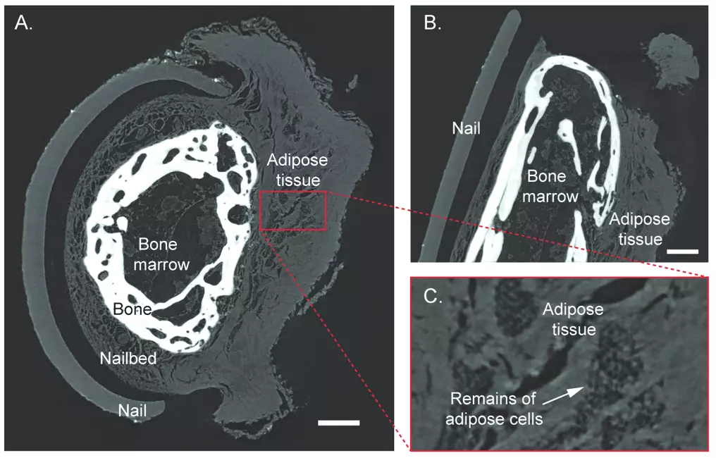
Mummies and other ancient remains are commonly examined by x-ray imaging due to the critical need for non-destructive techniques. Bone provides the strongest contrast and has been the main source of information. In paleopathology, the study of ancient diseases, significantly more information can be extracted if also studying the soft tissue at high resolution. Shown is a 2400-year-old ancient Egyptian mummy hand scanned with the Exciscope technology.

A. Axial view through tip of middle finger. B. Sagittal section. C. Adipose tissue with the remains of adipose cells. Scalebars represent 1 mm.
“Reproduced from J. Romell et al., “Soft-Tissue Imaging in a Human Mummy: Propagation-based Phase-Contrast CT”, Radiology (2018).”
Products & Services
Contact
William Twengström
X-ray Scientist
william.twengstrom@excillum.com
+46 76 022 36 15